


Study Identifies DNA Enhancers Involved in Hearing-Cell Regeneration
A study has identified DNA regulatory elements that help some animals, including fish and lizards, replace damaged hearing-related sensory cells. The discovery may guide future work on hearing and balance disorders, but does not show that human inner-ear cells can be regenerated.
The inner ear contains sensory cells that detect sound and supporting cells that provide their environment. In highly regenerative species, supporting cells can turn into replacement sensory cells after injury. Humans, mice, and other mammals generally lack this capacity. Scientists at the University of Southern California reported the stem-cell research in Proceedings of the National Academy of Sciences (PNAS). The work was led by Tuo Shi, Ksenia Gnedeva, and Gage Crump of USC's Keck School of Medicine.
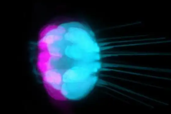
Figure caption. In the zebrafish inner ear, supporting cells shown in magenta produce new sensory hearing cells shown in blue. The study identifies DNA control elements important for supporting-cell hair-cell regeneration after injury in zebrafish, lizards, and other regenerative species. Image: Tuo Shi / Crump and Lozito labs / USC Stem Cell.
To understand how sensory-cell genes are reactivated in supporting cells, the researchers first examined genome organization in the sensory and supporting cells of regenerative zebrafish and green anoles. They then compared regulatory DNA for sensory genes with that of mice, which cannot replace inner-ear sensory cells after damage.
Crump, a USC professor of stem-cell biology and regenerative medicine, said comparing two regenerative vertebrates—fish and lizards—with nonregenerative mice revealed principles behind sensory-cell replacement. The team identified regulatory DNA elements called enhancers. After injury, these enhancers promote production of the protein ATOH1, which triggers expression of genes needed to form inner-ear sensory cells.
Using gene editing, the scientists deleted all five enhancers in zebrafish. Sensory-cell formation during development was impaired, as was regeneration after injury. Crump noted that deleting a single enhancer had often had little effect in earlier work, whereas targeting all five showed their collective importance.
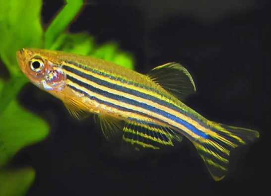
Zebrafish also have related sensory cells in the lateral line, an aquatic organ for detecting water flow and pressure. The five-enhancer deletions affected inner-ear sensory-cell formation and regeneration but not the lateral-line cells, pointing to a tissue-specific effect.
The researchers found similarly active enhancers in progenitors of mouse inner-ear sensory and supporting cells during embryonic development. In adult zebrafish and lizards, by contrast, these enhancers stay open in supporting cells, preserving their ability to replace injured sensory cells. Crump described the cells as “sister” populations with a common ancestor but different functions, retaining accessible enhancers from development into adulthood. He suggested that a future strategy might seek to open comparable enhancers in the human inner ear; that is a research possibility, not a demonstrated treatment.
Background: the organ of Corti
The organ of Corti, or spiral organ, is the hearing receptor on the basilar membrane of the cochlear duct. It contains inner and outer hair cells—the sensory cells discussed here—and several kinds of supporting cells, including pillar, phalangeal, and border cells. Hair cells convert mechanical movement caused by inner-ear fluid waves into neural signals sent along the auditory nerve to the brain's hearing pathways.
Generic image credit in the original article: online image, with no specific creator identified beyond the study figure credit above. Original ScienceDaily report.
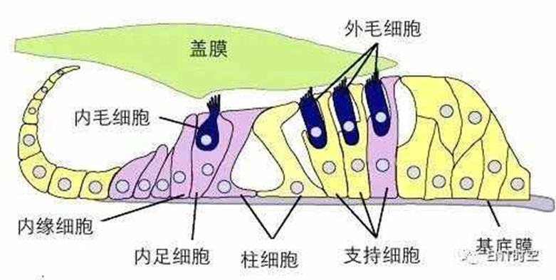
Labels identify the tectorial membrane, inner hair cell, inner border cells, inner phalangeal cells, pillar cells, outer hair cells, supporting cells, and basilar membrane.